Figure 2.
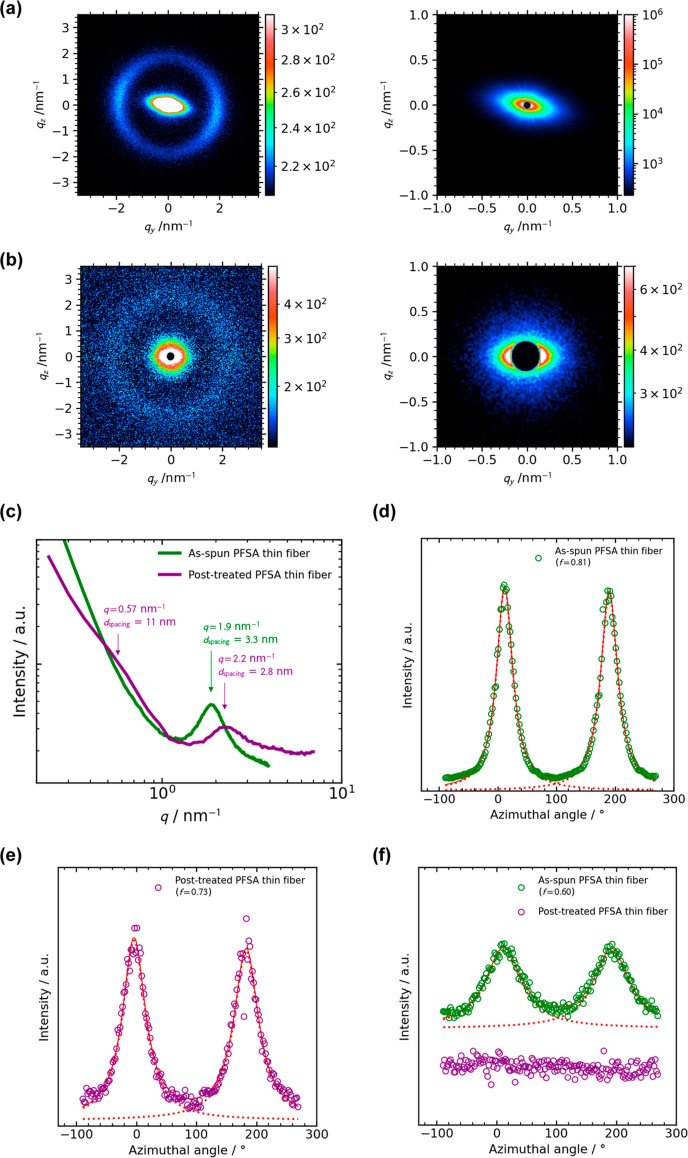
2D SAXS patterns of (a) the as-spun and (b) post-treated aligned PFSA thin fibers. Right-side images are the enlarged patterns of the left-side ones. The meridian direction of (a,b) is the fiber axis direction. (c) 1D profiles of (a,b). The azimuthal profiles of the matrix-knee at q = 0–1 nm–1 of the (d) as-spun and (f) post-treated aligned PFSA thin fibers. (e) The azimuthal profiles of the ionomer peak at q = 2–3 nm–1 of the as-spun and post-treated aligned PFSA thin fibers. All SAXS measurements were carried out at 25 ± 1 °C and 30–40% RH.

