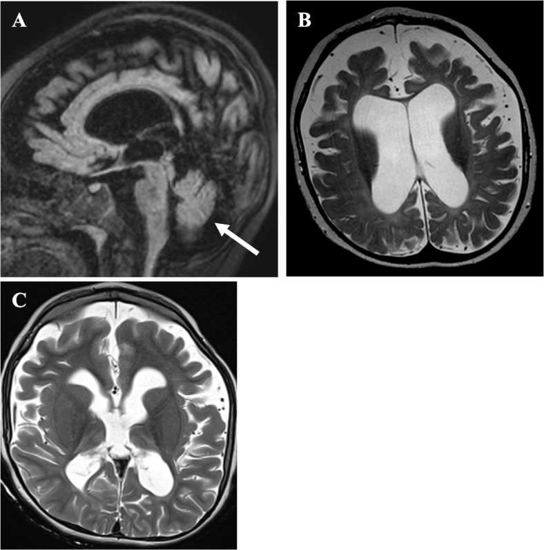
Fig. 1.

Brain MRIs of a patient with RARS2-related Mitochondrial Disorder. (Top Left) Sagittal FLAIR image of brain MRI at twelve months of age showing large subarachnoid spaces, ventriculomegaly, and generalized brain atrophy. There is a minimal reduction in brainstem size, and the volume of the cerebellum is preserved (white arrow). (Top Right) Axial T2W image at twelve months of age demonstrating ex vacuo enlargement of the ventricles and widening of the subarachnoid spaces. (Bottom Left) Axial T2W image at four years and six months of age demonstrating new T2 hyperintense signals in the bilateral thalami in addition to generalized volume loss, thinning of the corpus callosum, ex vacuo enlargement of the ventricles, and widening of the subarachnoid spaces
