
Figure 4: Clamping of the proximal femoral vessels using two separate microsurgical clamps.
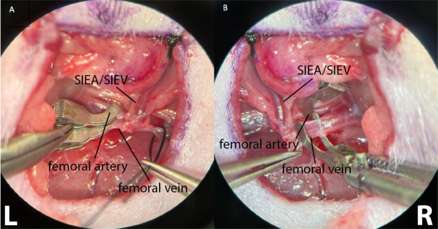
This allows better clamping control, ensuring the absence of arterial and retrograde venous flow. (A) shows both the left (L) femoral vessels clamped. The superficial inferior epigastric vessels are visible (SIEA/SIEV). (B) shows a clamped femoral artery and a femoral vein before clamping, on the right inguinal crease of the animal (R). Magnification: 40x.