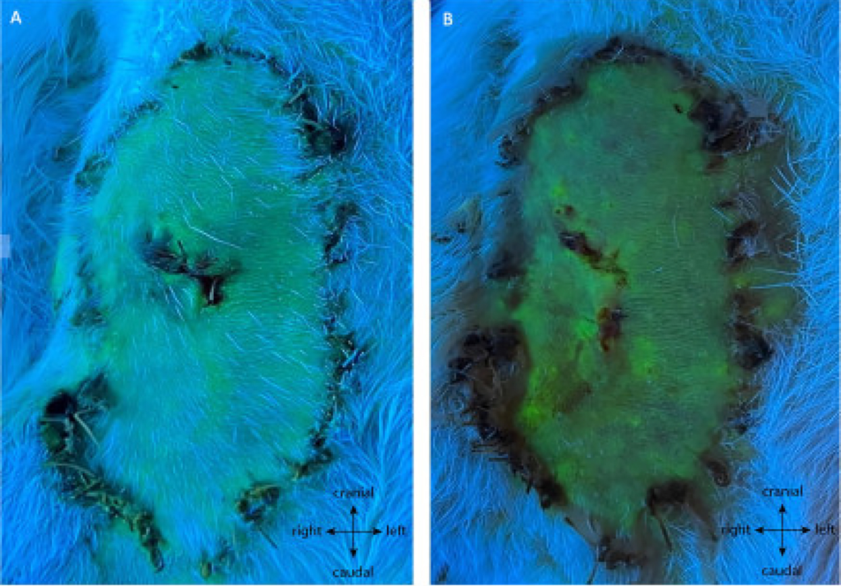
Figure 7: Angiography of the negative control at (A) POD5 and (B) POD10.

This assessment shows full survival of the flap without intervention on its pedicle. The green fluorescence shows well-perfused tissue, including the whole flap paddle. Note: control biopsies were taken on this replicate. Magnification: 40x.
