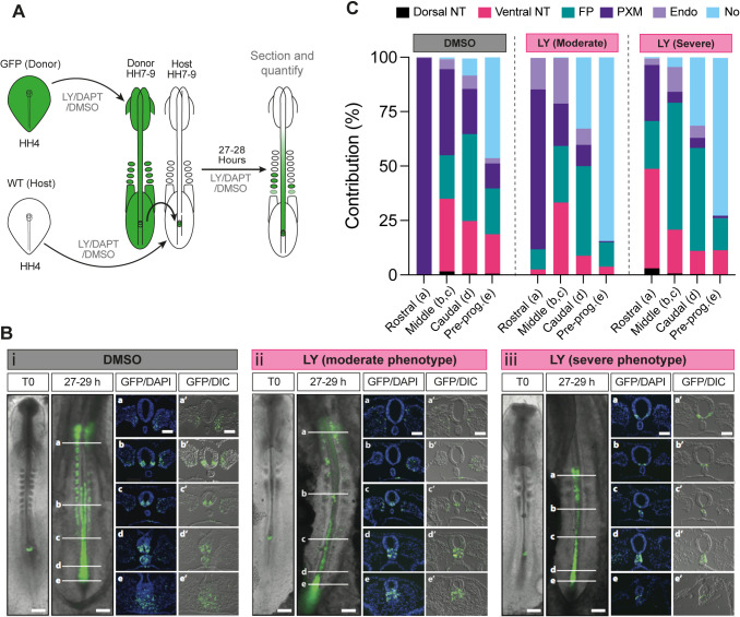
Fig. 4.
Notch signalling influences the contribution profile of axial progenitor cells in vivo. (A) Scheme depicting the experimental design/treatment regimens of chick embryo grafting experiments. WT, wild type. (B) Whole-mount embryo at the time of receiving an NSB graft (T0) and the GFP contribution pattern following culture in the presence of DMSO (i) or the Notch inhibitor LY in both the moderate (ii) and severe (iii) phenotype embryos after 27-29 h following the graft. Scale bars: 500 μm. Transverse sections at the level of the white indicator lines (a, b, c, d, e) show the nuclear stain DAPI and GFP or DIC with GFP (a′, b′, c′, d′, e′). Images are representative of independent experiments (analysed sectioned embryos: DMSO n=9, LY severe n=4/9 and moderate n=5/9). (C) Quantification of the proportion of GFP cells in transverse sections at position a (rostral), b and c (middle), d (caudal) and e (pre-progenitor, pre-prog.) contributing to axial and paraxial structures [dorsal neural tube (dorsal NT), ventral neural tube (ventral NT), floor plate (FP), paraxial mesoderm (somites rostrally and presomitic mesoderm caudally, PXM), endoderm (Endo) and the notochord (No)] in DMSO and LY-treated cultures.

