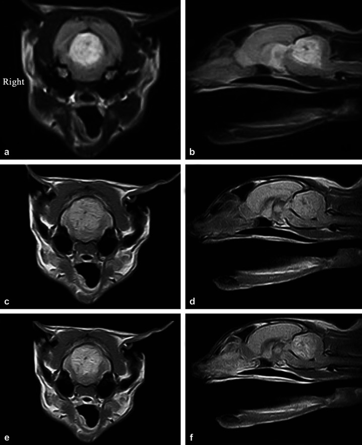
Fig 1.
(a) Transverse T2W image at the level of the cranial cerebellum showing a symmetrical, circular heterogeneously hyperintense lesion with ill-defined margins. (b) Sagittal T2W image showing a heterogeneously hyperintense area in the cranial two thirds of the cerebellum with some hypointense streaking. Note also the dorsal compression of the brainstem and slight protrusion of the cerebellum through the caudal foramen. (c) Transverse T1W image showing a mildly hyperintense lesion with some hypointense streaking when compared with the adjacent parenchyma. (d) Sagittal T1W image showing the cranial cerebellum to be mildly hyperintense compared with the surrounding parenchyma and including some hypointense patches. There is also mild cerebellar herniation through the foramen magnum and compression of the brainstem. (e) Post-gadolinium T1W image showing heterogenous enhancement. Within the lesion there are smaller isointense ring enhanced areas. (f) Post-gadolinium T1W sagittal image demonstrating heterogenous enhancement of the lesion with poorly defined margins.

