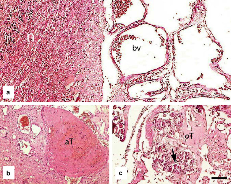
Fig 2.
(a) Photomicrograph showing normal cerebellum (n) adjacent to the large focal, expansive area containing multiple blood vessels (bv). (b) Photomicrograph of section through an acute thrombus (aT); several nearby blood vessels are abnormally thin-walled and dilated. (c) In some regions the thrombi had become organised (oT) and contained small deposits of mineralisation (arrow). Scale bar a–c: 75 μm.

