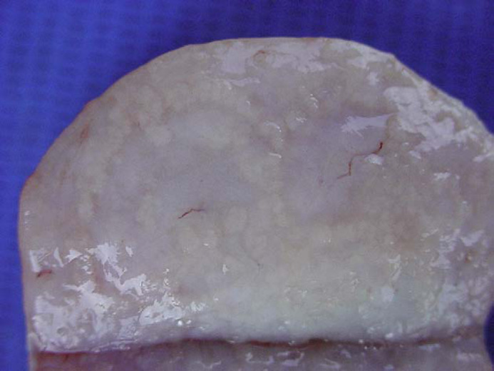
Fig 3.

Feline mammary fibroepithelial hyperplasia. Case 8. Mammary gland, cut surface. Multiple small, well-circumscribed, finely lobulated, contiguous, yellowish areas (corresponding to hyperplastic mammary ductal epithelium on histology) on a whitish, homogeneous, glistening background (that corresponds microscopically to proliferated interlobular stroma).
