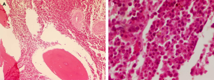
Fig 2.
Case 1 – histopathological section of a surgical biopsy of the lumbar vertebral body L6. There is extensive infiltration of inter-trabecular spaces by a neoplastic round cell population. The infiltrating cells have abundant eosinophilic cytoplasm with eccentrically located hyperchromatic round-to-oval nuclei consistent with plasmacytoid cells (haematoxylin and eosin stain, original magnification ×200 (A) and ×1000 (B)).

