Fig. 1.
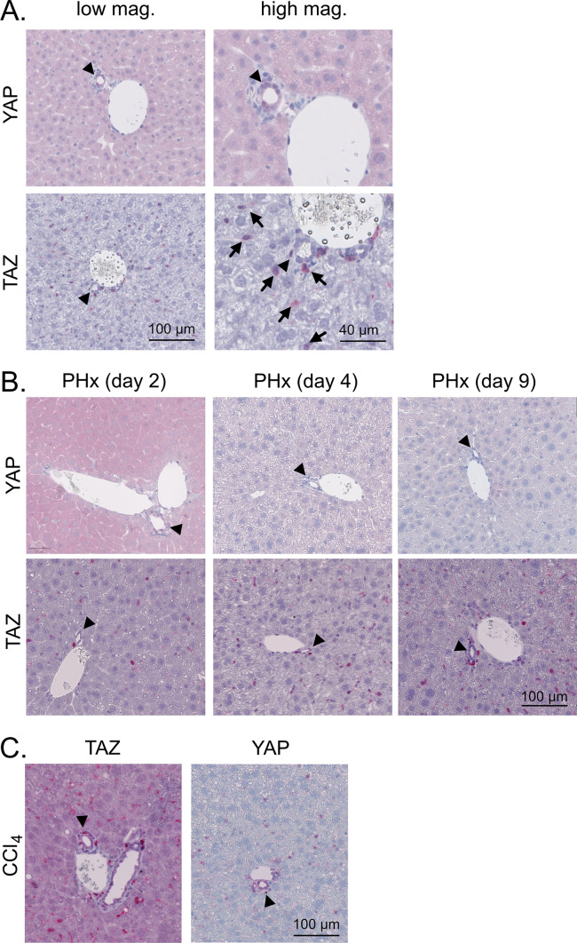
Hepatic expression of YAP and TAZ in healthy, regenerating, and fibrotic liver tissues. A Immunohistochemical stains of murine YAP and TAZ in healthy liver tissues of 10-week-old mice. Arrowheads: BECs/bile ducts, arrows: sinusoidal cells. B Staining of YAP and TAZ after 70% PHx. Samples were collected at different stages of hepatic regeneration: 2 days (proliferation), 4 days (reorganization), and 9 days (termination). Arrowheads: BECs/bile ducts. C Immunohistochemical stains of YAP and TAZ in CCl4-induced liver fibrosis. Ten-week-old mice were treated with CCl4 for six weeks followed by four weeks without injections. Arrowheads: BECs/bile ducts

