FIG. 1.
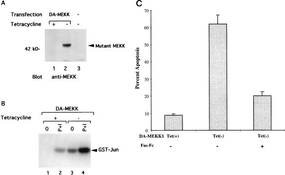
Inducible expression of DA-MEKK1 in Jurkat cells leads to constitutive JNK activation and induction of apoptosis. (A) Western blot showing the inducible expression of DA-MEKK1 in stably transfected Jurkat-tTA cells. Jurkat-tTA cells were transfected with 30 μg of cDNA encoding DA-MEKK1 in the pUHD10.3 vector (lanes 1 and 2). Cells in lane 3 were untransfected Jurkat-tTA cells. Following selection in 270 μg of hygromycin/ml for 4 weeks, the cells were grown in the presence (+) or absence (−) of 0.1 μg of tetracycline/ml for 24 h. Total cell lysates from 5 × 10⁶ cells were separated by SDS–10% PAGE and transferred to an Immobilon-P membrane. The membrane was overlaid with 0.1 μg of anti-MEKK1 antibody/ml, followed by HRP-conjugated protein A, and was developed by ECL. (B) In vitro kinase assay showing the constitutive activation of JNK by DA-MEKK1. The transfected cells described above were either left untreated (lanes 1 and 3) or stimulated for 10 min with 100 nM PMA and 1 μg of ionomycin/ml (P+I) at 37°C (lanes 2 and 4), and JNK activity was measured as previously described (14). (C) Cell viability assay in DA-MEKK1-expressing cells and the effect of Fas-Fc fusion protein. DA-MEKK1 Jurkat cells were incubated in the presence or absence of 30 μg of Fas-Fc/ml and grown under tet+ or tet− conditions for 72 h. Cell viability was measured by trypan blue exclusion. Duplicate counts were performed by two independent observers. We have previously shown that in these cells, trypan blue uptake is accompanied by 7AAD uptake and DNA laddering (14). Similar results were obtained in three separate experiments.

