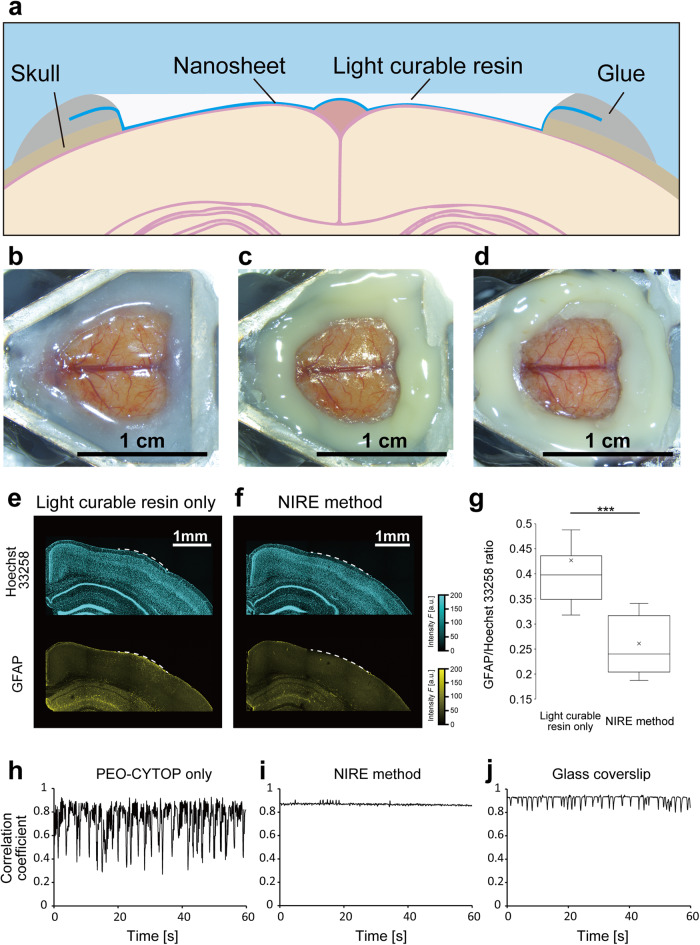
Fig. 1. Construction of cranial windows from PEO-CYTOP nanosheets and light-curable resin.
a Fixing the light curable resin on the PEO-CYTOP nanosheet. Schematic illustration of the ‘nanosheet improved by light-curable resin (NIRE)’ method. b Exposure of the dura surface by craniectomy. c Sealing the brain surface with a PEO-CYTOP nanosheet. d Fixing light-curable resin on the PEO-CYTOP nanosheet. e Immunostaining of astrocytes using anti-GFAP and nuclear counterstaining in a brain slice obtained 4 weeks after craniectomy and covering with light-curable resin only (without a PEO-CYTOP nanosheet). The white dashed line indicates the region where the skull was removed, and the resin was fixed. f Immunostaining of astrocytes using anti-GFAP and nuclear counterstaining in a brain slice obtained 4 weeks after craniectomy and the NIRE method. The white dashed line indicates the region where the skull was removed, and the resin was fixed. g The ratio of mean GFAP to Hoechst 33258 signal per 300 × 300 µm2 (15 ROIs from three mice in each condition). ***p < 0.005 by Welch’s t test. h Time series of a correlation coefficient calculated from two-photon images of SR101-labeled astrocytes acquired through a cranial window consisting of a PEO-CYTOP nanosheet in the awake condition. i Time series of a correlation coefficient calculated from two-photon images of SR101-labeled astrocytes acquired through a cranial window produced using the NIRE method in the awake condition. j Time series of a correlation coefficient calculated from two-photon images of SR101-labeled astrocytes acquired through a cranial window using the glass coverslip method in the awake condition.

