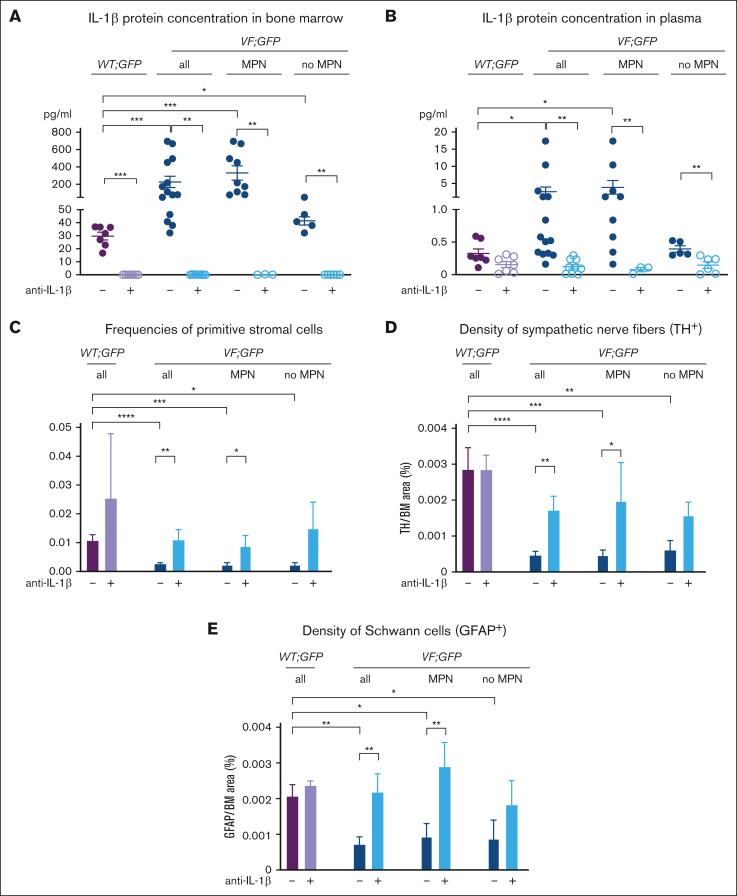
Figure 5.
Inhibition of IL-1β preserves the HSC niche in the BM. (A) IL-1β protein concentration in BM lavage (1 femur and 1 tibia) and (B) plasma of WT mice (from supplemental Figure 8), and JAK2-V617F mice (from Fig. 4) with or without MPN phenotype. Nonparametric Mann-Whitney 2-tailed t test was performed for statistical comparisons. The lower limit of detection was 0.11 pg/ml. (C) Frequency of primitive stromal cells (CD45−CD31−Ter119−Sca1−PDGFRα+) in the skull of mice transplanted with BM from WT;GFP or VF;GFP donor mouse after 18 weeks of treatment with isotype or anti–IL-1β antibody (from Figure 4). Two-tailed unpaired t tests were performed for statistical comparisons. (D) Bar graphs show the quantification of tyrosine hydroxylase (TH) area and (E) of glial fibrillary acidic protein (GFAP) area in the BM. One-way analysis of variance with uncorrected Fisher least significant difference test was performed for statistical comparisons. All data are presented as mean ± SEM; ∗P < .05; ∗∗P < .01; ∗∗∗P < .001; and ∗∗∗∗P < .0001.

