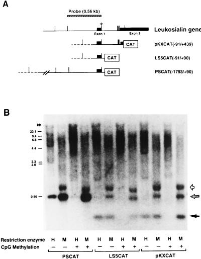
FIG. 2.
Methylation states of the 5′ regions of the leukosialin gene exogenously introduced and stably maintained in non-leukosialin-expressing HeLa cells. (A) Schematic representations of the leukosialin gene and leukosialin CAT constructs. The exons are depicted by filled boxes, and introns and the 5′ flanking regions are depicted with horizontal lines. The vector sequences are depicted with dotted lines. MspI (CCGG) sites are shown with vertical bars. The asterisk indicates the polymorphic site, where an MspI site is lost in one allele of HeLa cells. The MspI DNA fragment (560 bp) used for a hybridization probe is presented at the top. (B) Genomic DNAs (10 μg) from HeLa cells stably transfected with CpG-methylated- or unmethylated-leukosialin CAT constructs were digested with HpaII (H) or MspI (M) and separated by 1.5% agarose gel electrophoresis. The blotted filter was hybridized with the 560-bp DNA fragment of the 5′ region of the leukosialin gene shown in panel A. The hatched arrow indicates the position of the fragments produced by the endogenous gene as well as the exogenously introduced gene. The filled arrow indicates the signal produced by the exogenously introduced gene. The open arrow indicates the position of a polymorphic fragment of the endogenous leukosialin gene of HeLa cells.

