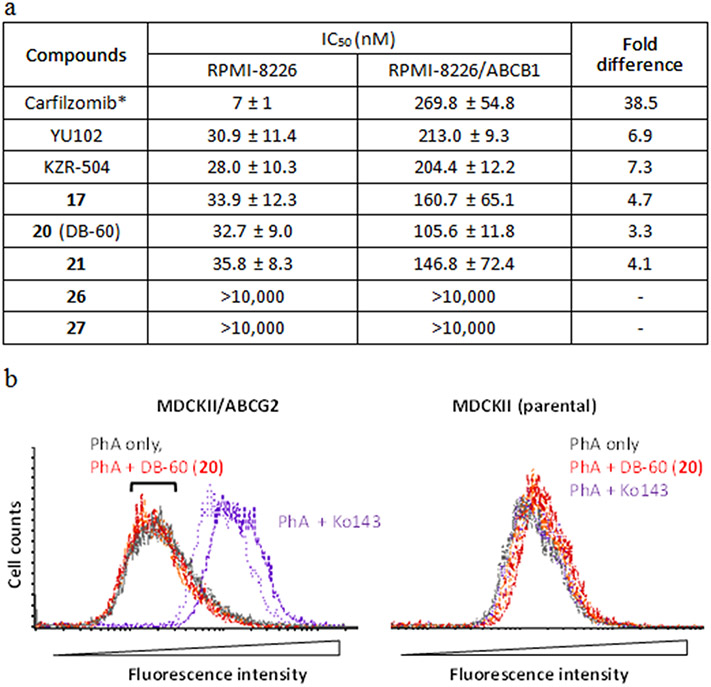
FIGURE 3.
(a) LMP2 inhibitory activity of DB-60 (20) and other compounds in ABCB1-overexpressing RPMI-8226 cells. ABCB1-overexpressing RPMI-8226 cells or the parental RPMI-8226 cells were incubated with varying concentrations of respective compounds (n=3 replicates per concentration). After 72 hours, cell lyses were prepared to measure proteasome catalytic subunit activities (LMP2 and LMP7). Curve fitting analysis was performed to calculate IC50 values reported as the mean ± SD. *Due to carfilzomib's cytotoxic effect (a dual inhibitor of cP and iP), cells were incubated for 3 hours, followed by the measurement of chymotrypsin-like (CT-L) activity. (b) Lack of interactions between DB-60 (20) and ABCG2. The inhibitory interactions with ABCG2 were assessed by measuring the changes in the cellular accumulation of pheophorbide A (PhA, a fluorescent ABCG2 substrate) using MDCKII cells stably expressing ABCG2 (established in our previous study41) and parental cells. The known ABCG2 inhibitor Ko143 was used as a positive control. After cells were preincubated in the complete medium containing PhA (1 μM) with Ko143 (0.2 μM) or DB-60 (20) (2 or 5 μM) at 37°C for 30 min, cells were then washed with ice-cold medium and incubated again with Ko143 or DB-60 (20) for 45 min at 37°C. Subsequently, the fluorescent signal was measured via flow cytometry using the excitation and emission wavelengths at 635 and 670 nm, respectively. Colored lines represent the following groups: grey, PhA only; orange, PhA in the presence of 20 (2 μM); red, PhA in the presence of 20 (5 μM); purple, PhA in the presence of Ko143 (0.2 μM) (n=2 per group).

