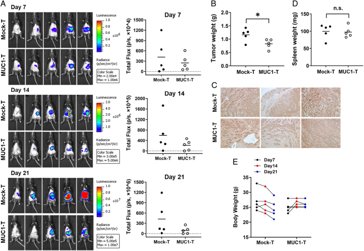
FIGURE 5.
MUC1 CAR T cells reduce KCM-Luc tumor growth in the pancreas. Mouse pancreatic cell line KCM-luciferase (20,000 cells/mouse) was orthotopically injected into the pancreas of MUC1.Tg mice. Seven days postsurgical injection, the presence of a tumor in the pancreas was confirmed by IVIS imaging. On day 8, mice were randomized and received a single i.v. injection of mock T cells as control, or MUC1 CAR T cells. A, Tumor growth was monitored by weekly IVIS imaging. On the right panels, the total flux of bioluminescence was quantified by marking a region of interest covering the tumor xenograft. B, The tumor wet weight for individual mice on day 21 (endpoint; P = 0.0353). C, IHC staining for tumor MUC1 with TAB004 (×100). Brown staining shows tMUC1 positivity (n = 3), three mice from each group were used for sectioning and staining. D, The spleen wet weight of individual mouse at the endpoint. E, The body weight change before and after T-cell treatment. No significance is observed between the mock-T group and the MUC1 T-cell group. n = 5 mice per group. The horizontal bar marks the mean value in each group. Two experiments are conducted with one representative data being shown here. *P <0.05 (unpaired t test). CAR indicates chimeric antigen receptor; i.p., intraperitoneal; i.v., intravenous; MUC1, mucin 1; tMUCI, tumor form of mucin 1.

