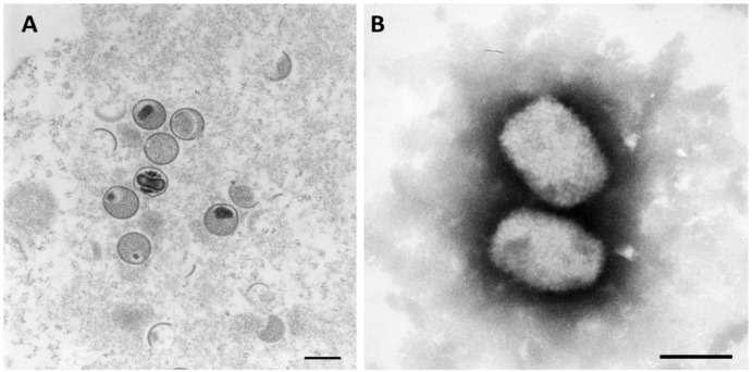
Figure 3.
Monkeypox virus under transmission electron microscope. (A) utilizing ultrathin stained sections; Bar = 200 nm, (B) utilizing negative stained; Bar = 200 nm. *mature: oval-shaped virus particles, immature: crescents and spherical shape. Source: Robert Koch Institute (https://www.rki.de) 4 September 2022 [61].

