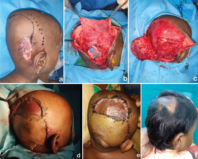
Fig. 3.

( a ) Preoperative marking showing incision ( thick line ) for rotation flap and a dotted line showing the temporoparietal fascial (TPF) flap marking. ( b ) The TPF flap was raised through the same incision. ( c ) Flap transposed onto the implant and sutured as a snuggly fitting pocket. ( d ) The traditional rotation flap was raised along the same incision with split-thickness skin graft (STSG) for donor area. ( e ) Immediate post-op. ( f ) Late post-op.
