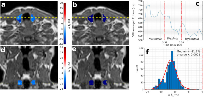
Fig. 3.
Example OE-MRI parametrical maps of thyroid gland (patient number 5). T1 weighted VIBE image is shown with overlaid parametric ΔT1 map of the thyroid gland (a, d, axial and coronal plane respectively) and overlaid statistical map of ΔT1 times (b, e). Blue colour indicates statistically significant decrease in T1 times, white indicates no statistically significant change, and red indicates statistically significant increasing T1 times. c Time series of T1 times averaged over the entire thyroid VOI. f Histogram of ΔT1 times for the entire thyroid VOI. OE-MRI Oxygen-enhanced magnetic resonance imaging, VIBE Volumetric interpolated breath-hold examination, VOI Volume of interest

