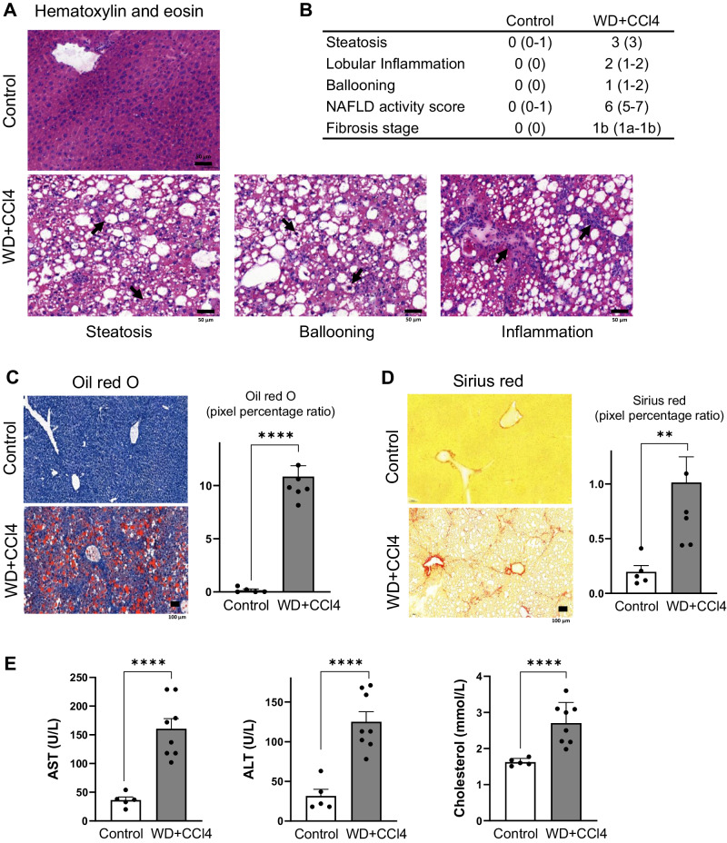
Fig. 1. Effect of WD + CCl4 on mouse liver.
A Microscopy of hematoxylin and eosin (H&E)-stained liver sections showing diffuse macro-vesicular steatosis, lobular inflammation, and the presence of ballooned hepatocytes in the WD+CCl4 group. B Histological scoring of the WD+CCl4 group livers, given as median values. Values in parentheses denote the range of values across all mice. C Oil Red O staining comparing neutral lipid content of control and WD+CCl4 group (unpaired t-test; ****p < 0.0001). D Microscopy of Sirius Red-stained liver sections showed increased fibrosis in WD+CCl4 group (unpaired t-test; **p < 0.005). E Liver transaminases (ALT and AST), and total cholesterol were significantly increased in WD+CCl4 group (unpaired t-test; ****p < 0.0001). All box plot error bars represent standard deviation. WD+CCl4: Western diet supplemented by carbon tetrachloride. (WD+CCl4 n = 7, control n = 5).

