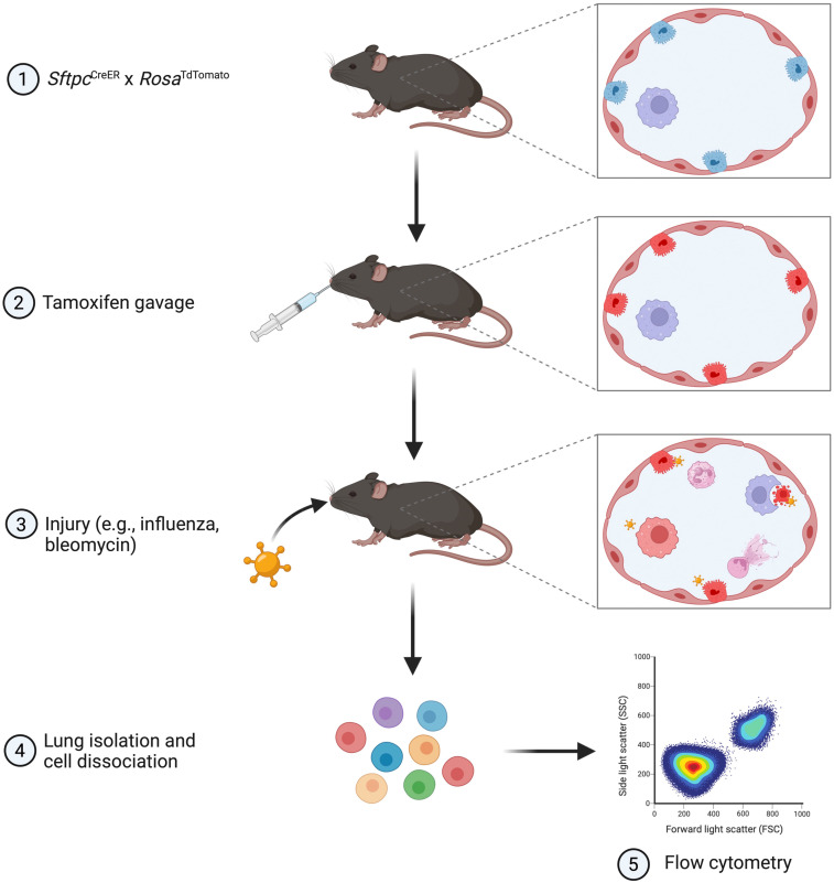
Figure 1.
Schematic of the workflow for the in vivo efferocytosis model. The SftpcCreER × RosaTdTomato mice were given tamoxifen to label alveolar type 2 (AT2) cells with red fluorescence. Mice were then injured with intranasal influenza or bleomycin to induce AT2 cell apoptosis. At the appropriate time point, murine lungs were isolated and dissociated into single-cell suspensions for immunostaining and analysis by flow cytometry. This figure was created with BioRender.com.

