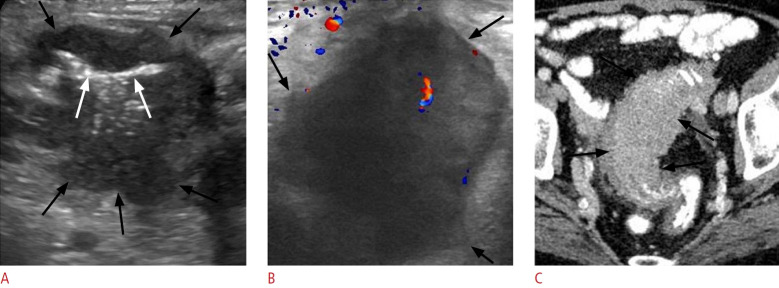
Fig. 10. Sigmoid cancer in a 69-year-old man.
A. Transverse sonographic image at the left lower quadrant depicts asymmetric mural thickening of the sigmoid colon (black arrows) with hypoechoic echogenicity. This finding is characterized by an irregular and lobulated contour, along with narrowing of the lumen (white arrows). B. Sonographic image taken with a high-frequency linear-array transducer at a more distal level reveals the irregular contour of the lesion as well as prominent lobulations (black arrows) and evidence of internal vascularity. C. Corresponding axial contrast-enhanced computed tomography image confirms segmental wall thickening of the sigmoid colon (black arrows), featuring a lobulated contour and luminal narrowing.

