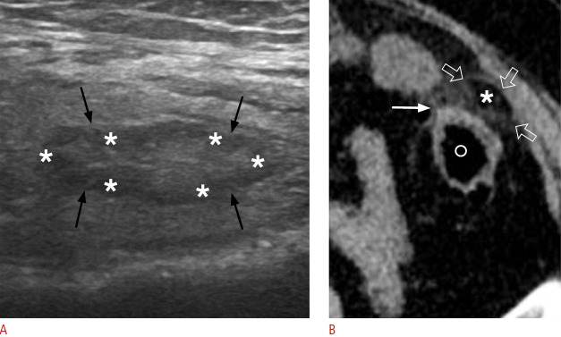
Fig. 12. Epiploic appendagitis in a 58-year-old woman.
A. Sonographic image at the site of maximal tenderness in the left lower quadrant reveals an ovoid, hyperechoic lesion (black arrows) surrounded by a hypoechoic halo (asterisks). B. Corresponding axial non-enhanced computed tomography image displays a fat-density ovoid structure (asterisk) adjacent to the descending colon (labeled with "o"), surrounded by a thin hyperdense rim (open arrows) and perilesional inflammatory fat stranding (white arrow).

