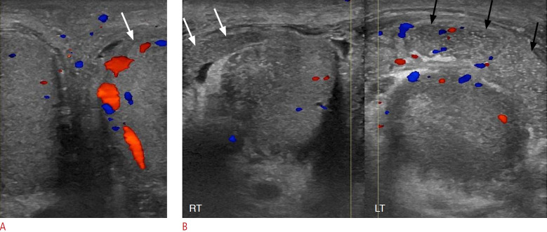
Fig. 14. Acute left epididymo-orchitis in a 56-year-old man.
A. Sonographic side-by-side comparison of the right and left testes shows increased vascularity in the left testis (arrow) compared to the normal contralateral testis. B. Sonographic side-by-side comparison of the right and left epididymides shows enlargement, coarse echotexture, and hypervascularity of the left epididymis (black arrows) compared with the right epididymis (white arrows).

