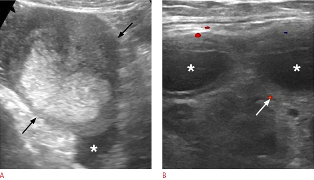
Fig. 20. Ovarian torsion in a 34-year-old woman.
A. Sonographic image of the left lower quadrant reveals a markedly enlarged left ovary with heterogeneous internal echogenicity (black arrows) and free fluid in the cul-de-sac (asterisk). B. A high-frequency transducer provided a focused image of the lesion, displaying peripherally located follicles (asterisks) and minimal stromal vascularity (white arrow), with an arterial waveform present (not shown). Venous flow was not detected.

