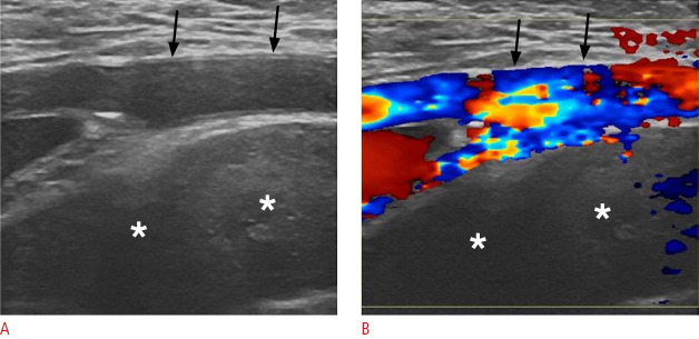
Fig. 23. Spontaneous retroperitoneal hemorrhage in a 65-year-old man.
A, B. Sonographic images of the left lower quadrant reveal an extensive heterogeneous fluid collection (asterisks), without internal vascularity, situated deep to the external iliac artery (black arrows). This location is sonographically accessible for the evaluation of the retroperitoneum.

