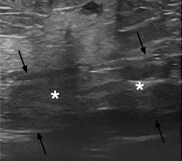
Fig. 24. Rectus sheath hematoma in a 37-year-old woman on anticoagulant therapy.

Sonographic image of the anterior abdominal wall reveals an enlarged left rectus abdominis muscle (black arrows) with a heterogeneous echotexture (asterisks) consistent with a hematoma.
