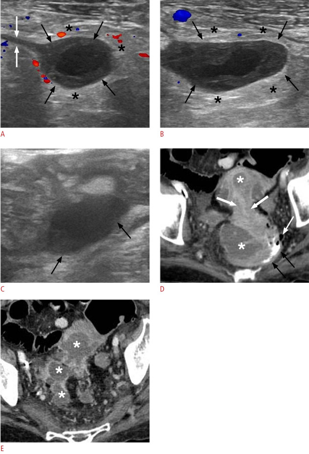
Fig. 4. Acute complicated (Hinchey stage II) colonic diverticulitis in a 70-yearold woman.
A-C. Pelvic ultrasonographic images reveal ovoid, thick-walled fluid collections (black arrows indicative of pericolic and distant [A and B] or intramural [C] abscesses). These are encircled by a hyperechoic halo (asterisks in A and B), which corresponds to perilesional inflammatory fat changes. A fistulous tract is visible as a linear hypoechoic structure (white arrows in A), establishing a connection between the collection and the colonic lumen (not shown). D, E. Axial contrastenhanced computed tomography images demonstrate extensive irregular wall thickening with hypoattenuating submucosa (thick white arrows in D) and pronounced mucosal enhancement, indicative of colonic inflammation. Multiple fluid-filled collections with rim enhancement are present along the affected sigmoidal segment (asterisks) or at distant intrapelvic sites, consistent with abscesses. A small amount of contrast is observed within the lumen of the distal sigmoid (black arrows in D), accompanied by diverticula (thin white arrow).

