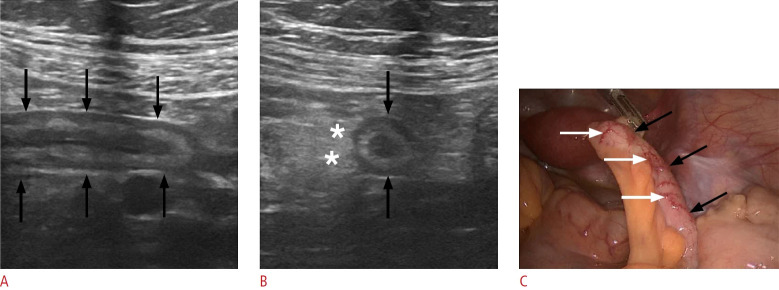
Fig. 5. Acute appendicitis in a 14-year-old woman.
A. Sonographic image of the right lower quadrant displays the blind-ending appendix along its long axis (black arrows), with an outer diameter of 9 mm and preserved stratification. B. A sonographic image through the short axis of the appendix reveals appendiceal enlargement (black arrows) and periappendiceal hyperechoic, inflammatory fat changes (asterisks). C. A laparoscopic intraoperative image of the same patient exhibits a dilated appendix (black arrows) with mural hyperemia (white arrows).

