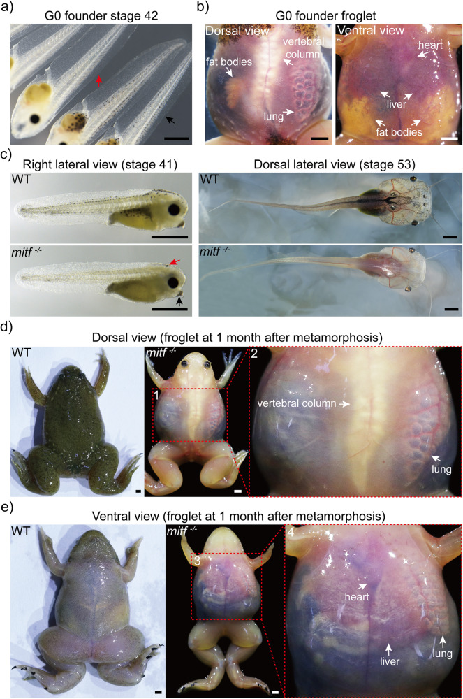
Fig. 1. Biallelic mitf disruption makes Xenopus tropicalis transparent within two months post-metamorphosis.
a After using the CRISPR/Cas9 system to knock out mitf via gRNA T7, founder generation (G0) mosaic tadpoles at stage 42 exhibited a loss of melanophores (indicated by red arrow), compared to wild type (WT) tadpoles from the same batch (indicated by black arrow). 79 mosaic tadpoles and 20 wild type tadpoles were observed. b One month after metamorphosis, the transparent skin of the mosaic froglet allowed for external visibility of internal organs such as the fat bodies, lung, liver, heart, and vertebral column. Here was shown one representative froglet, out of a total of 11 mosaic froglets. c The F1 mitf−/− Xenopus tropicalis tadpole exhibited melanophores loss throughout the entire body at stages 41 and 53 compared to wild type tadpoles from the same batch. Notably, at stage 41, the melanin pigmentation in the eyes of mitf−/− Xenopus tropicalis was generated by retinal pigment epithelium (RPE) cells, and redistribution of melanin in oocytes resulted in some melanin pigment in the head (indicated by red arrow) and cement gland (indicated by black arrow). However, the melanin pigment in the head and cement gland disappeared in later development stages, such as stage 53. Representative photographs were exhibited from 35 F1 mitf−/− Xenopus tropicalis tadpoles at stage 41, 15 F1 mitf−/− Xenopus tropicalis tadpoles at stage 53, as well as from 10 wild type tadpoles each at stage 41 and 53. d, e One month after metamorphosis, the F1 mitf−/− Xenopus tropicalis froglet exhibited transparent skin from both dorsal (d) and ventral (e) views. Red dashed boxes 2 and 4 corresponded to magnified views of red dashed boxes 1 and 3, respectively. The transparent skin allowed for external visibility of internal organs including the lung, liver, heart, and vertebral column within one month after metamorphosis as shown. Ten F1 mitf−/− Xenopus tropicalis froglets and ten wild-type Xenopus tropicalis froglets were used to provide representative photographs as shown. Each scale bar is 1 mm.

