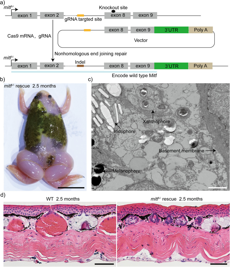
Fig. 5. Restoration of the damaged mitf site in mitf−/−Xenopus tropicalis results in the rescue of the phenotype of melanophores, xanthophores, and granular glands.
a The precise integration of the DNA fragment encompassing the final two exons and the 3′UTR region of mitf into the mitf−/− genomic locus via CRISPR/Cas9-mediated non-homologous end joining repair has been demonstrated. b A rescued phenotype was demonstrated in a representative 2.5-month-old froglet of the mosaic founders. Approximately 300 embryos of mitf−/− Xenopus tropicalis were injected, and eventually, six tadpoles were found to have a rescued phenotype of melaphores. c TEM examination of the dorsal skin of the rescued froglet depicted in b revealed a significant resurgence of both xanthophores and melanophores. We sampled three distinct regions of the dorsal skin. Six ultrathin sections were subsequently generated from each of the three samples to yield the representative results presented here. d The histological structure of the dorsal skin from 2.5-month-old WT froglets and rescued froglets was presented. Three 2.5-month-old WT froglets and three rescued froglets depicted in b were subjected to H&E staining, and two dorsal skin samples with 3 mm×3 mm were obtained from each frog. Histological examination was performed on ten paraffin sections for each collected sample to generate the representative results presented here. Scale bar in b, 1 cm; Scale bar in c, 1 μm; Scale bar in d, 50 μm.

