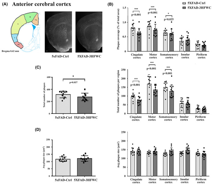
FIGURE 3.

3HFWC treatment reduces the plaque load in the anterior cerebral cortex of 5XFAD female mice. (A) Representative section showing approximate location of cortical regions quantified separately on three serial coronal brain sections (left) and representative photomicrographs of Thioflavin S‐positive amyloid deposits in 4‐month‐old female 5XFAD‐Ctrl and 5XFAD‐3HFWC mice (right). Scale bar = 500 μm. (B) Quantification plot of the percent area covered by Thioflavin S‐positive amyloid plaques in cortical subregions of control (5XFAD‐Ctrl, light gray bars) or mice treated with 3HFWC substance (5XFAD‐3HFWC, dark gray bars). (C) The total plaque numbers in three sections per mouse were counted and are indicated as the number of plaques in the whole cortex (left) or in cortical subregions (right). (D) Average plaque size (μm2) in three sections/mouse in the whole cortex (left) or in cortical subregions (right). In all panels, data are shown as mean ± SD (n = 10 mice per group). Statistical significance was analyzed by Mann–Whitney U test as the composite (average) histological score from several sections of an individual mouse.
