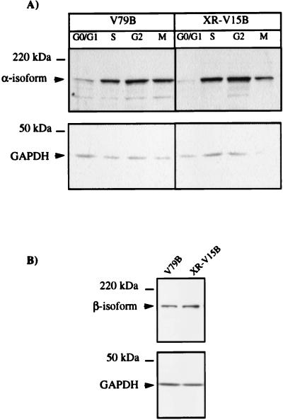
FIG. 4.
Wild-type and Ku86-deficient cells show similar levels of α and β-isoforms of DNA topoisomerase II. (A) Whole-cell extracts were obtained from G1/G0-, S-, G2-, and M-phase cells (as described in Materials and Methods), and 75-μg aliquots of proteins of the different extracts were analyzed by Western blotting using a polyclonal antibody directed against DNA topoisomerase IIα. (B) A total of 5 × 10⁵ exponentially growing cells were lysed with 1% SDS at 90°C for 5 min and loaded in each lane. DNA topoisomerase IIβ levels were analyzed by using a monoclonal antibody (3H10) directed against this isoform. The lower part of the membranes was probed with an anti-GAPDH antibody to ensure that comparable amounts of protein were loaded in all lanes. Representative results of three assays are shown.

