Figure 2.

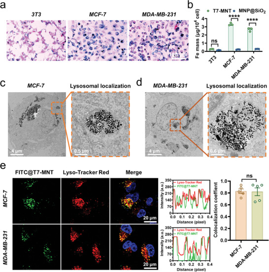
Lysosome targeting ability of T7‐MNTs in cancer cells. a) Prussian blue staining of T7‐MNTs co‐incubated with three different cell lines (3T3, MCF‐7, and MDA‐MB‐231 cells) for 24 h (scale bar: 100 µm). b) The internalized Fe mass in 3T3, MCF‐7, and MDA‐MB‐231 cells after co‐incubation with T7‐MNTs and MNP@SiO2, respectively (n = 3, ****p < 0.0001, ns: no significance). c,d) Bio‐TEM images of T7‐MNTs located in lysosomes inside MCF‐7 c) and MDA‐MB‐231 cells d) (scale bar: 4 µm, 0.5 µm). e) Confocal fluorescence images and colocalization coefficient of FITC@T7‐MNTs with lysosomes in MCF‐7 and MDA‐MB‐231 cells (scale bar: 20 µm). The representative line profiles of FITC@T7‐MNTs and Lyso‐Tracker Red (red line in images) were measured by Image J (n = 5, ns: no significance).
