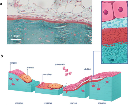
Figure 6.

Alternative aOs/osteoblasts interface and schematic illustration of typical bone remodeling with the proposed mechanism for twisted plywood formation through acidic collagen mesophase. a) Light microscopy overview of a bone histological section stained with Goldner trichrome showing that nOs is not always seen; osteoblasts can be in contact with aOs. b) The schematic representation of the proposed mechanism for bone remodelling process includes the different steps observed here and matching observations described in the literature. (right) After the activation phase, bone resorption occurs via osteoclasts (multinucleate cell) through proteolytic enzymes and protons release (resorption phase). After further debris removal via macrophages, osteoblasts (mononuclear cuboid cells in pink) produce collagen fibrils (forming the newly osteoid tissue in light blue) (reverse phase) that progressively dissolve into or interact with the acidic ECM domains (in red) in which a collagen‐based mesophase is formed via molecule accretion. Thereafter, stabilization of the cholesteric geometry occurs through the co‐precipitation of collagen fibrils; MB is formed exhibiting a mineralized twisted plywood pattern (formation phase). (left) Schematic representation of the collagen molecules‐based domain (aOs) that co‐exists between two fibrillar tissues (nOs and MB).
