FIG. 1.

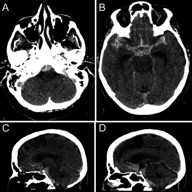
Head CT without contrast obtained during initial presentation, showing extensive subarachnoid hemorrhage within the premedullary (A), prepontine (C and D), suprasellar, and bilateral sylvian (B) cisterns.

Head CT without contrast obtained during initial presentation, showing extensive subarachnoid hemorrhage within the premedullary (A), prepontine (C and D), suprasellar, and bilateral sylvian (B) cisterns.