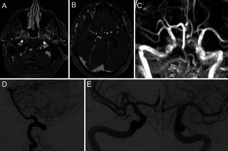
FIG. 2.
Initial vascular imaging. MR angiography (A) showing a right vertebral artery dissecting aneurysm. No additional vascular abnormalities were seen in the anterior circulation on time of flight (B) or maximum intensity projection (MIP; C) sequences. Diagnostic right vertebral artery angiogram (D), lateral view, with an arrow pointing to the dissecting aneurysm in the vertebral artery just proximal to the posterior inferior cerebellar artery. Right and left internal carotid artery injection angiograms (E), anteroposterior view, showing no additional vascular abnormalities.

