FIGURE 1:
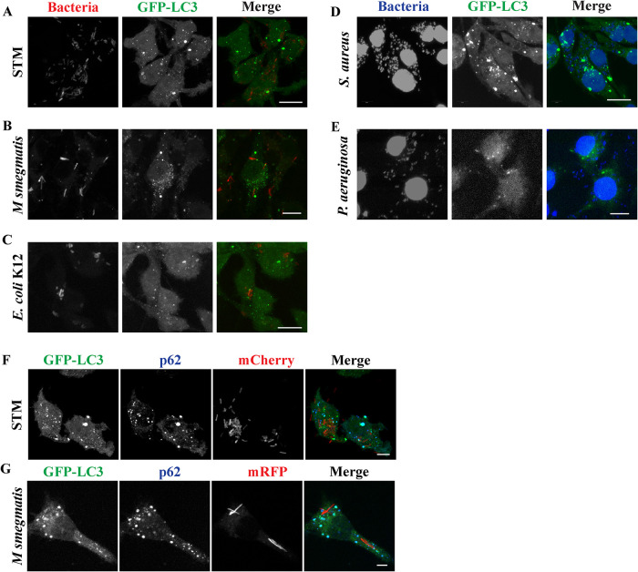
Formation of GFP-LC3 (green) dots in RAW264.7 macrophages. RAW264.7 cells stably expressing GFP-LC3 form GFP-LC3 positive puncta after 6 to 12 h post infection by STM-mCherry (A), Mycobacterium smegmatis-mRFP (B), E. coli K12-mCherry (C), DAPI stained strains of Staphylococcus aureus (D), and Pseudomonas aeruginosa (E). RAW 264.7 cells form ALIS positive for GFP-LC3 (green) and p62 (blue) in response to infection with STM-mCherry (F) or M. smegmatis-mRFP (G). Scale bars, 5μm.

