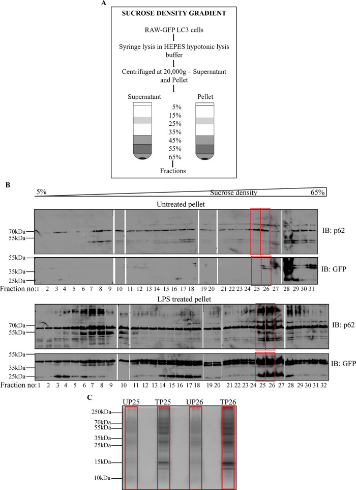
FIGURE 5:
Schematic to represent fractionation of cell lysates by SDG centrifugation (A). Immunoblotting of untreated and LPS treated pellet fractions for GFP and p62 (B). Coomassie stained SDS–PAGE gel indicates an enrichment of proteins in treated fractions as compared with untreated fractions (C).

