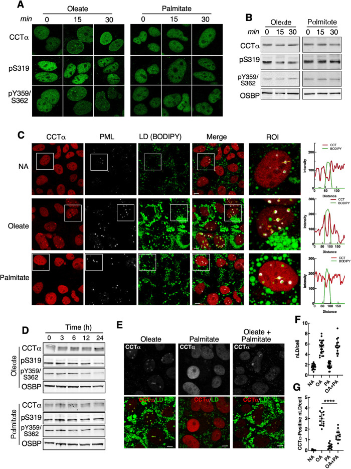
FIGURE 4:
LAPS and nLDs formed by palmitate treatment do not recruit CCTα. (A) Huh7 cells treated with oleate (0.4 mM) or palmitate (0.4 mM) for 0, 15, and 30 min were immunostained with antibodies against CCTα, CCTα-pS319, and CCTα-pY359/pS362. (B) immunoblot of lysates from cells treated as described in Panel A. (C) Huh7 cells treated with no addition (NA), oleate (0.4 mM), or palmitate (0.4 mM) for 24 h were immunostained for CCTα and PML, and LDs were visualized with BODIPY493/503 (bar, 5 μm). RGB line plots for CCTα and BODIPY are for selected LAPs (yellow line) in the region of interest (ROI) panel. (D) immunoblot analysis of CCTα phosphorylation in Huh7 cells treated with oleate or palmitate for up to 24 h. (E) immunostaining of Huh7 cells that were treated with oleate (0.4 mM), palmitate (0.4 mM) or oleate plus palmitate (0.3 and 0.1 mM, respectively) for 24 h (bar, 5 μm). (F) quantification of nLDs in cells treated with no addition (NA), oleate (0.4 mM, OA), palmitate (0.4 mM, PA), oleate and palmitate (0.4 and 0.1 mM) for 24 h. (G) quantification of CCTα-positive nLDs as described in panel F. Results in panels F and G are from three experiments that quantified 12–16 images (12–16 cells/field). Significance was determined using a Students t test; ****p < 0.0001.

