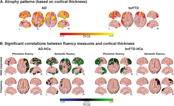
FIGURE 4.

Surface‐based morphometry results. (A) Atrophy of persons with AD and bvFTD, relative to healthy controls. Reduced cortical thickness was observed predominantly in temporo‐parietal areas for AD and frontotemporal regions for bvFTD. (B) Associations between significant fluency variables and whole‐brain cortical thickness. Valid responses in the phonemic and semantic tasks were positively correlated with cortical thickness in various brain regions in the AD‐HC and bvFTD‐HC groups. In the AD‐HC group, valid responses in the phonemic task were positively correlated with cortical thickness in the left putamen, middle and posterior cingulate gyri, calcarine fissure, lingual gyrus, and right rolandic operculum, while valid responses in the semantic task were positively correlated with thickness in the left middle temporal gyrus and the right superior temporal and middle cingulate gyri. In the bvFTD‐HC group, valid responses in the phonemic task were positively correlated with thickness in the left anterior cingulate, precentral gyrus, middle occipital lobe, and calcarine fissure, as well as the right middle superior frontal gyrus, the right parahippocampus, and the bilateral posterior cingulate gyrus and precuneus. Frequency in the semantic task was linked to cortical thickness in both AD‐HC and the bvFTD‐HC groups, involving temporo‐parietal and cingulate cortices in the AD‐HC analysis, and frontotemporal and cingulate cortices in the bvFTD‐HC analysis. AD, Alzheimer's dementia; bvFTD, behavioral variant frontotemporal dementia; HC, healthy control; I, inferior; L, left; R, right; S, superior; TFCE, threshold‐free cluster enhancement
