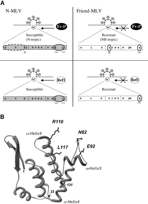
FIG. 4.
Schematic representation of the Fv1 and Ref1 restriction targets. (A) Target and masking determinants for Fv1b (top) and Ref1 (bottom) restrictions in the MLV CA. Key residues involved in the susceptibility of N-MLV to Fv1b and Ref1 restriction genes are circled (left panels, R110 and L117). The dashed circle indicates a fragment found to modulate the restriction level (see text). Residues sufficient to make the Friend MLV CA target inaccessible to Fv1b and/or Ref1 restrictions are circled (right panels, combined D92 and H117 for Fv1b; H117 for Ref1). (B) Three-dimensional structure of the amino-terminal domain of N-Akv CA (adapted from reference 21). Amino-terminal (position 1) and carboxy-terminal (position 131) extremities of the crystallized fragment are indicated as well as position 33 corresponding to the BamHI cloning site shown in Fig. 1 and α-helices 4, 5, and 6. Representation of asparagine 82 (N82), aspartate 92 (E92), arginine 110 (R110), and leucine 117 (L117) side chains highlights their potential accessibility to restriction factors.

