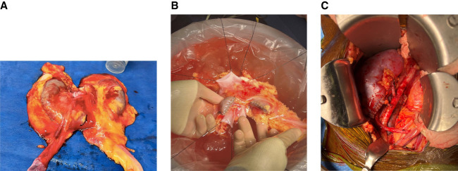
Figure 1.
Horseshoe kidney transplantation. (A) Horseshoe kidney with each moiety about 9 cm in size and fused at inferior poles by a 3-cm wide isthmus. (B) Horseshoe kidney moieties were separated by dividing the isthmus using a 60-mm blue gastrointestinal anastomosis stapler and oversewn using a 5–0 chromic gut suture. (C) Immediately post-transplant, each horseshoe kidney moiety reperfused well with no bleeding at the divided site.

