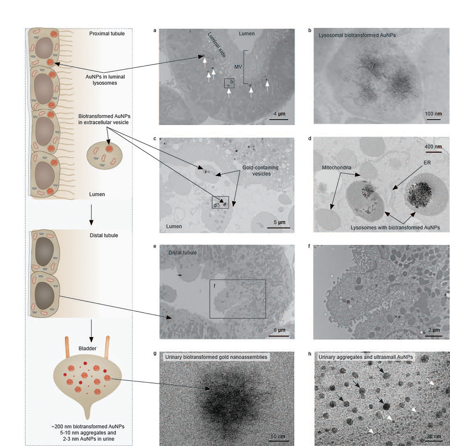
Fig. 3 |. Biotransformation and re-excretion of endocytosed (+)-AuNPs by PTs.

a, A representative EM image of PTs at 24 h p.i. of (+)-AuNPs showing the intracellular (+)-AuNPs located in lysosomes of PTECs on the luminal side (labelled by arrows). The labelled box is the area shown in b. b, A representative EM image of the biotransformed 200–300 nm flower-like gold nanoassemblies composed of nanofibres in a lysosome of PTEC. c, A representative EM image of extracellular vesicles in a proximal tubular lumen that contain lysosome-encapsulated biotransformed AuNPs and other organelles. d, A magnified image of the extracellular vesicle in c containing lysosome-encapsulated biotransformed AuNPs, mitochondria and smooth ER. e,f, Representative EM images of distal tubules showing that no biotransformed AuNPs were found inside the distal tubules at 24 h p.i. g,h, Representative EM images of biotransformed gold nanostructures, including ~200 nm gold nanoassemblies (g) and 5–10 nm AuNPs (h, indicated by black arrows), found in the urine within 24 h p.i. of (+)-AuNPs, in addition to ultrasmall AuNPs with an original size of 2–3 nm in the urine (h, indicated by white arrows). No gold or silver enhancement staining was used for EM samples. Representative EM images in a, c, e, g and h are presented out of images acquired from three independent samples. The image along the left side shows a schematic of the process.
