
Fig. 6 |. Nanoparticle endocytosis and organelle extrusion significantly reduced in PTs with cisplatin-induced injury.
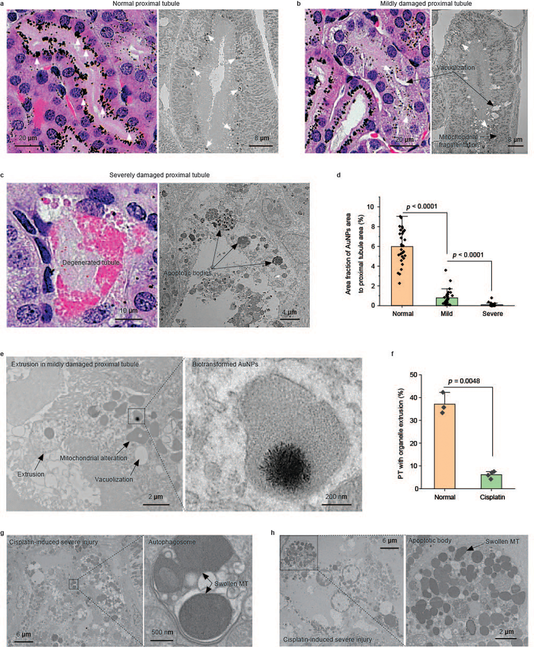
a–c, Representative pathological and EM images of the PTs in cisplatin-injected mice showing three distinct injury statuses. a, A normal-appearing PT with no obvious structural damage in the pathology and no ultrastructural damage in the EM image. A high uptake of (+)-AuNPs was observed (AuNPs are indicated by white arrows). b, A mildly damaged PT. Intracellular vacuolization is clearly identified in the pathology. Both vacuolization and mitochondrial fragmentation are observed in the EM image. Less uptake of (+)-AuNPs was observed (AuNPs are indicated by white arrows). c, A severely damaged PT. Degenerated proximal tubular structure is observed. Consistently, cell apoptosis with apoptotic bodies is observed in the EM image. Almost no (+)-AuNPs are observed in the severely damaged PTs. d, Quantification of the area fraction of silver-enhanced AuNPs in the entire cross-section of a PT in normal-appearing, mildly damaged and severely damaged PTs. N = 30, 30 and 32 proximal tubular cross-sections in H&E-stained kidney tissue slides were analysed for normal, mildly damaged and severely damaged PTs, respectively. Data are presented as mean ± s.d. P = 5.16 × 10⁻¹⁸ between normal and mild. P = 3.75 × 10⁻⁵ between mild and severe. Two-sided Student’s t-test was performed at the 0.05 significance level. e, Representative EM images of the extrusion and biotransformation of lysosomal AuNPs in PTs with a mild injury. The biotransformation of AuNPs was unaffected in mildly damaged PTs. f, Percentages of PTs with organelle extrusion in normal mice and cisplatin-treated mice after (+)-AuNP injection showing that tubular organelle extrusion was significantly reduced in acute tubular injury. N = 3 tissue sections from three individual tissue blocks, mean ± s.d. More than 70 proximal tubular cross-sections were analysed for each tissue block. Two-sided Student’s t-test was performed at the 0.05 significance level. g,h, Representative EM images of an autophagosome and apoptotic body in cisplatin-induced severely damaged PTs. No gold or silver enhancement staining was used for EM samples.