
FIG. 6.
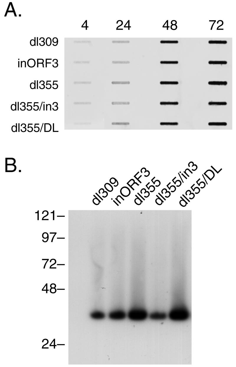
Viral DNA accumulation and concatenation in GM07166 (Nbs1−) cells. (A) GM07166 cells (Nbs1−) were infected with wild-type or mutant viruses at a multiplicity of 200 particles per cell. Total nuclear DNA was isolated at the times indicated and diluted (1:5, 4 h; 1:10, 24 h; 1:100, 48 h; 1:100, 72 h) and then applied to a nylon membrane by using a slot blot apparatus. The blot was hybridized with a fluorescently labeled probe corresponding to the left end of the Ad5 genome. The signal was detected and quantified by using a Molecular Dynamics Storm 860 PhosphorImager and ImageQuant software. (B) GM07166 cells were infected with wild-type or mutant viruses at a multiplicity of 200 particles per cell. Cells were harvested at 72 h postinfection, and nuclear DNA was prepared for PFGE as described in Materials and Methods. Following electrophoresis, the DNA was transferred to a nylon membrane, probed with a 32P-labeled Ad total genome probe, and visualized by autoradiography. Molecular weight standards (in kilobase pairs) are indicated on the left.