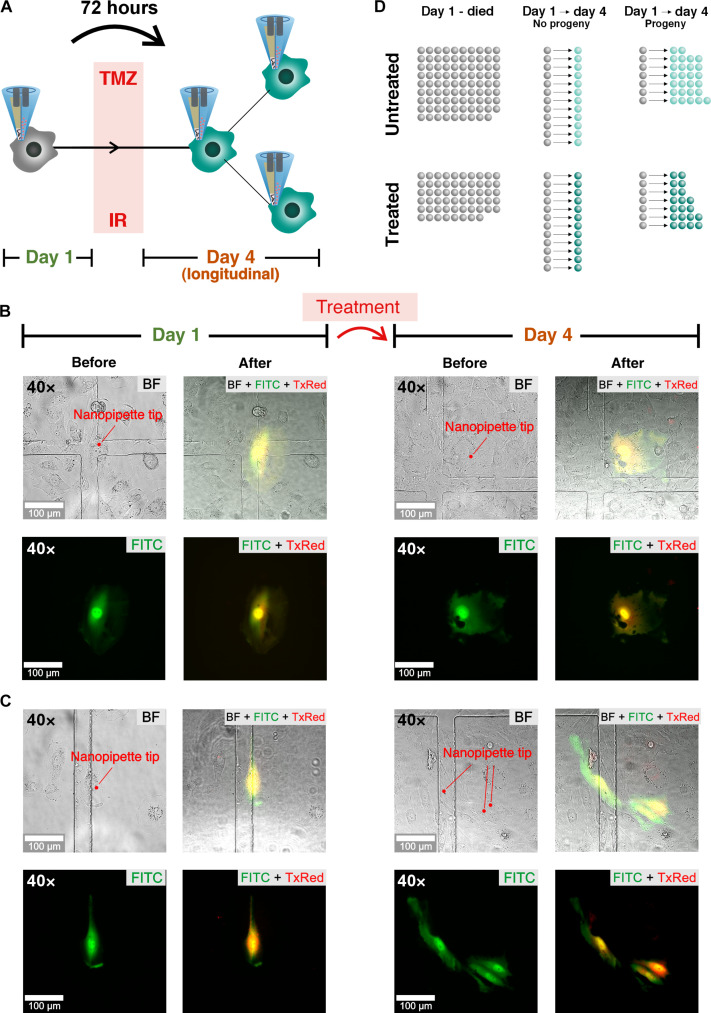
Fig. 3. Longitudinal nanobiopsy and nanoinjection of individual glioblastoma cells through therapy.
(A) An individual GBM cell undergoes nanobiopsy and nanoinjection on day 1. Following standard treatment of temozolomide TMZ and irradiation (IR) the same cell or its progeny undergoes a second nanobiopsy and nanoinjection on day 4 (longitudinal), 72 hours after the first nanobiopsy. (B) Optical (BF) and fluorescence (FITC, TxRed) micrographs of an individual M059KGFP cell before and after the first (day 1) and longitudinal nanobiopsy following standard treatment (day 4). (C) Optical and fluorescence micrographs of an individual M059KGFP cell undergoing initial nanobiopsy and nanoinjection (day 1), surviving treatment and dividing, and subsequent longitudinal nanobiopsy and nanoinjection of its progeny following treatment (day 4). (D) Illustration of the nanobiopsy count of untreated and treated cells that were nanobiopsied on day 1 and died (day 1–died), nanobiopsied on day 1, survived, did not divide, and nanobiopsied again on day 4 (day 1 and day 4–no progeny) and cells that were nanobiopsied on day 1, divided, and whose progeny was nanobiopsied again on day 4 (day 1 and day 4–progeny).

