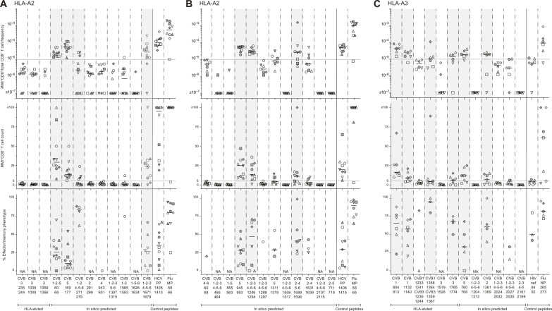
Fig. 2. Screening of CVB peptides for recognition by blood CD8+ T cells in CVB-seropositive healthy adults.
(A to C) HLA-A2–restricted (A and B) and HLA-A3–restricted candidates (C) were tested with combinatorial MMr assays [see reproducibility in fig. S2 (F and G)]. Each symbol represents a donor (legends in table S1), and the bars display median values. For each panel, the top graph depicts the frequency of MMr+CD8+ T cells out of total CD8+ T cells, with the horizontal line indicating the 10−5 frequency cutoff used as a first validation criterion; the middle graph displays the number of MMr+CD8+ T cells counted, with the horizontal line indicating the cutoff of five cells used to assign an effector/memory phenotype; the bottom graph shows the percent fraction of effector/memory cells (i.e., excluding naïve CD45RA+CCR7+ cells) among MMr+CD8+ T cells (for those donors with ≥5 cells counted; NA when not assigned). Peptides validated in this screening phase are highlighted in gray. In each panel, control peptides are derived from viruses eliciting predominantly naïve responses in these unexposed individuals (HLA-A2–restricted HCV PP1406–1415 and HLA-A3–restricted HIV nef84–92) and from influenza virus (Flu MP58–66 and NP265–273 peptides) eliciting predominantly effector/memory responses.

