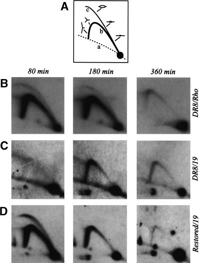
Figure 2.
The ori-β′ locus in the DR8 cell line is inactivated in early S phase. (A) Principle of the neutral/neutral 2D gel replicon mapping method (Brewer and Fangman 1987), where curve a denotes the arc of linear fragments, curve b the single fork arc, and curve c the bubble arc. Cells were synchronized at the G1/S boundary by release from an early G1 block into mimosine for 12 h. After drug removal and return to drug-free medium, samples were taken in early, mid-, and late S phase (80, 180, and 360 min, respectively), replication intermediates were prepared using EcoRI to digest the DNA, and the digests were separated on a 2D agarose gel. The DNA was then transferred to a membrane and hybridized with appropriate probes. (B) Replication pattern of the early-firing rhodopsin control origin in DR8 cells. (C) The transfer in B was stripped and rehybridized with a probe specific for the ori-β′ locus. (D) Replication pattern of the cell line obtained by restoring the deletion in DR8 by homologous recombination with cosmid KZ381.

