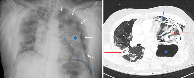
Figure 1.
Left panel: Four centimetres left-sided pneumothorax (white arrows) with associated left-sided lung atelectasis (asterisk) and pneumomediastinum (blue arrowheads) that appeared on day 14. Discrete right-sided mediastinal shift. Right panel: CT scan on day 15 showing a large left basal pneumatocele with an undrained fluid component (blue star). A chest tube was in the antero-medial position (blue arrow). There is densification of pulmonary infiltrate as previously described (red arrows).

